“Will removing my upper molar affect my sinus?” It is a reasonable question, especially when an X-ray shows the roots sitting close to the sinus.
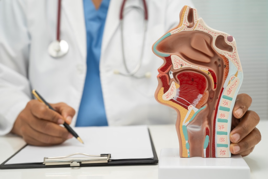
The maxillary sinuses are air-filled spaces located above the upper back teeth. In some people, the bone separating a tooth root from the sinus is very thin. Removing the tooth can occasionally create an opening between the mouth and sinus, known as an oroantral communication. Careful assessment before surgery helps us understand the anatomy and plan the extraction accordingly.
Why Upper Molar Extractions Require Careful Planning
The roots of the upper first, second, and third molars can lie close to the floor of the maxillary sinus. Root length, bone thickness, infection, and previous dental problems all influence the complexity of an extraction. Research has examined these anatomical factors and their relationship to oroantral communication.
This does not mean every upper molar extraction carries a high risk of sinus complications. It means the surgeon needs to assess the individual case rather than assume the anatomy is straightforward.
How We Assess Sinus-Related Risks Before Extraction
We assess the tooth’s position and proximity to the sinus to determine the safest extraction approach.
Reviewing X-rays and Additional Imaging
We begin by examining the tooth, reviewing the patient’s dental history, and assessing the available X-rays. These images help us evaluate root shape, surrounding bone, infection, and the tooth’s relationship to the sinus.
When conventional imaging doesn’t provide enough information for a complex case, we may consider cone-beam computed tomography (CBCT). It produces three-dimensional images that can clarify the relationship between the roots and the sinus floor. CBCT is not necessary for every extraction; the decision depends on the clinical findings. Research on CBCT and oroantral communication discusses its role in assessing upper third molar extractions.
Choosing a Controlled Surgical Technique
Once we understand the anatomy, we can plan the removal to minimize unnecessary trauma to the surrounding bone and tissue.
Depending on the case, the surgeon may divide the tooth into smaller sections rather than attempt to remove it in one piece. This can give better control during removal. The technique depends on the tooth’s condition, root shape, and position. No method eliminates every risk, but a planned approach helps avoid unnecessary force near the sinus.
What Happens If a Sinus Opening Is Suspected?
After removing the tooth, the surgeon assesses the socket and surrounding tissue. If the team suspects an opening between the mouth and sinus, it determines the next step based on its size, tissue condition, and whether infection is present.
Management may include stabilizing the site, placing sutures or protective material, and arranging follow-up. Some small openings can close with appropriate care, while larger or persistent communications may require surgical closure. The decision should follow a clinical assessment rather than the appearance of the socket alone.
Patients should not try to test for an opening by blowing air through the nose or repeatedly checking the extraction site. These actions can interfere with healing.
Sinus Perforation Symptoms to Watch For
Contact your oral surgeon if you notice any of the following after an upper molar extraction:
- Liquid passing from your mouth into your nose when drinking.
- An unusual sensation of air moving between your mouth and nose.
- Persistent drainage, an unpleasant taste, or a bad odor.
- New or worsening sinus pressure or one-sided nasal symptoms.
- Pain that does not improve as expected.
These symptoms alone do not confirm sinus perforation. Your surgeon can examine the area and decide whether you need further assessment or treatment.
Aftercare Instructions That Help Protect Healing
If your surgeon identifies a sinus-related risk or complication, follow the specific precautions provided for your case. These commonly include:
- Avoid blowing your nose.
- Sneeze with your mouth open rather than suppressing the sneeze.
- Avoid straws, smoking, vaping, and forceful spitting.
- Avoid strenuous activity during the period your surgeon recommends.
- Take prescribed medication exactly as directed.
- Attend scheduled follow-up appointments.
Pressure changes can place stress on the healing area. Do not stop precautions simply because the extraction site feels comfortable; follow your surgeon’s instructions about when normal activities can resume.
Planning Your Upper Molar Extraction
At Ridge Oral Surgery, patients can discuss their treatment needs, imaging findings, and recovery questions with the surgical team. If you need an upper molar removed and are concerned about its proximity to the sinus, schedule an evaluation with Ridge Oral Surgery.
Sinus perforation after tooth extraction is a recognized complication, but it does not occur with every upper molar removal. Reviewing the anatomy, choosing the right technique, and following postoperative instructions help ensure a well-managed procedure. If you notice unusual nasal symptoms after surgery, contact your surgeon promptly rather than attempting to assess the opening yourself.