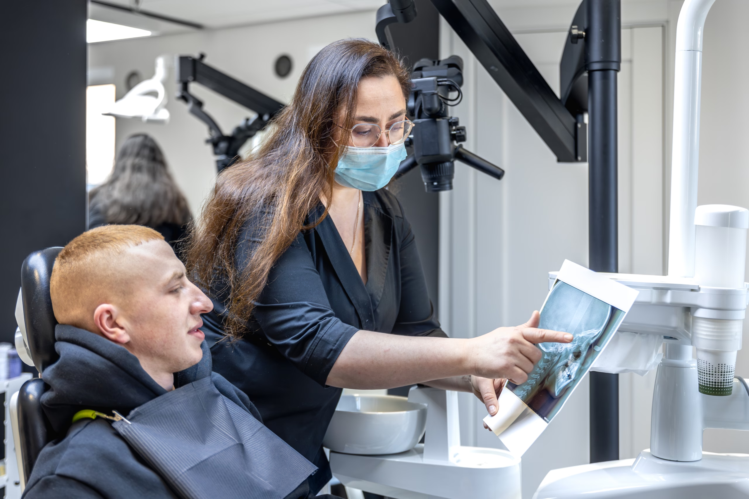
A normal X-ray doesn’t mean nothing is wrong. Standard dental films are two-dimensional pictures of a three-dimensional structure, and they routinely miss cracks, early bone changes, and anything hidden behind overlapping roots or bone.
Six things commonly cause genuine tooth pain with a clean film. Most are diagnosable — usually with better clinical testing, sometimes with better imaging.
Why a 2D X-ray misses things
Everything is flattened onto one plane. Roots overlap each other and overlap bone. A lesion sitting behind a root can be invisible from the front.
Bone loss has to reach a threshold before it shows. Early inflammation at a root tip doesn’t produce a visible dark area until enough mineral has gone. CBCT imaging detects around 17% more teeth with apical bone loss than intraoral radiographs do.
Cracks are almost impossible to see. A fracture line only shows on a conventional film when the X-ray beam happens to run nearly parallel to the fracture plane — within a few degrees. Miss that angle, and the crack is invisible. Conventional radiography is considered inconclusive for detecting vertical root fractures.
Soft tissue problems don’t show at all. Muscle, joint and nerve pain produce no radiographic change whatsoever.
So “the X-ray looks fine” means one specific thing: no change large enough and well-angled enough to appear on that image. It doesn’t mean your pain isn’t real, and it isn’t a verdict.
The six usual explanations
| Cause | The tell |
|---|---|
| Cracked tooth syndrome | Sharp pain on releasing a bite, not on pressing down. Often one specific cusp. Sensitivity to cold |
| Early pulpitis | Lingering pain after cold, more than 30 seconds. Worse lying down. May throb at night |
| Vertical root fracture | Usually a root-canal-treated tooth. Dull ache, a gum boil that comes and goes, deep narrow pocket |
| Sinus origin | Several upper back teeth ache at once. Worse leaning forward. Congestion, recent cold |
| Referred pain from jaw muscles | Worse on waking, worse after chewing. Clicking or jaw stiffness. Not pinpointable |
| Persistent dentoalveolar pain | Constant ache that didn’t resolve after a root canal or extraction. No clinical findings |
Cracked tooth syndrome
The most common answer, and the hardest to see. A crack can run through the crown and into the dentine without any radiographic sign at all.
The classic symptom is pain on release — you bite down on something, and the pain hits as you let go. Each chew flexes the crack slightly and irritates the pulp inside.
It’s diagnosed by biting on individual cusps one at a time, by transillumination, and sometimes by removing an existing filling to look directly. Not by an X-ray.
Early pulpitis
Inflammation inside the tooth, before it has produced any change in the surrounding bone. Reversible pulpitis settles if the cause is removed. Irreversible pulpitis doesn’t, and needs root canal treatment or extraction.
The useful distinction: brief sensitivity to cold that stops immediately is usually reversible. Pain that lingers more than 30 seconds after the cold is removed suggests the pulp is beyond recovery.
Vertical root fracture
Typically affects root-canal-treated teeth, particularly those with posts. The symptoms are vague — a dull persistent ache, a recurring gum boil, a deep isolated pocket beside the tooth.
This is where imaging makes a measurable difference. A meta-analysis found pooled sensitivity for detecting vertical root fractures of 0.71 for CBCT versus 0.52 for periapical radiography, with no significant difference in specificity. CBCT also distinguishes the two patterns: a cracked tooth typically shows intact cortical bone with angular defects near the fissure, while a vertical root fracture shows J-shaped defects, probing depths greater than 6 mm, and cortical bone loss.
Sinus origin
The roots of upper back teeth sit close to, sometimes within, the sinus floor. Sinus inflammation makes several of them ache simultaneously.
The pattern that gives it away: more than one tooth, worse when you lean forward or go down stairs, often with congestion or a recent cold, and no single tooth that reproduces the pain when tapped.
Referred pain from jaw muscles
Referred pain is a prominent feature of temporomandibular disorders, and the teeth are one of its regular destinations. People in this group often arrive having already had a root canal that didn’t help.
This guide to telling TMJ pain from tooth pain covers the distinguishing tests.
Persistent dentoalveolar pain
Sometimes called atypical odontalgia. Continuous pain in a tooth or extraction site with no identifiable pathology, often following previous dental treatment. It presents a genuine diagnostic challenge, and the most important thing about it is recognising it — because the wrong answer is more dental treatment on a healthy tooth.
A few others worth ruling out
- Recent dental work sitting high. A filling or crown fractionally too tall produces pain on biting that resolves in minutes once adjusted.
- Exposed root surface from gum recession — sensitivity rather than true toothache.
- Grinding and clenching, which makes teeth tender to bite on generally rather than at one point.
- An erupting or impacted wisdom tooth pressing on the tooth in front.
- Sinus-adjacent infection after an upper extraction, which needs separate assessment.
Rarely, pain in the jaw with exertion, breathlessness or chest discomfort can be cardiac. That’s an emergency department, not a dental appointment.
What better imaging adds, and when it’s worth it
A CBCT scan produces a three-dimensional volume rather than a flattened image, which removes superimposition and shows bone changes a flat film can’t resolve. For suspected root fractures, hidden lesions, sinus involvement and complex anatomy, it answers questions standard films can’t.
But it shouldn’t be routine. CBCT delivers a higher radiation dose than conventional dental radiography, and professional guidance is consistent that it’s a problem-solving tool rather than a screening tool. The right question is “what would this scan change about my treatment?” If there’s a clear answer, it’s indicated. If there isn’t, it isn’t.
Worth knowing: a scan that finds nothing is still useful. It rules out the structural causes and redirects attention to the muscular, neuropathic and sinus explanations — which is exactly what people who’ve had three unhelpful treatments need.
What else should happen before more treatment
A thorough workup for unexplained tooth pain includes:
- Cold testing, comparing against neighbouring and opposite-side teeth
- Bite testing on individual cusps, not the whole tooth
- Percussion, tapping each tooth
- Periodontal probing, looking for an isolated deep narrow pocket
- Transillumination, shining light through the tooth to reveal cracks
- Removing an existing filling to inspect the floor of the cavity directly
- Selective anesthesia — numbing one tooth at a time to localise the source
- Checking the bite for a high restoration
Selective anesthesia is particularly useful when you can’t say which tooth hurts. If numbing the suspect tooth doesn’t stop the pain, the source is elsewhere.
What to do next
- Go back and say it hasn’t resolved. “The X-ray was clear” is a starting point, not a discharge.
- Describe the pain precisely. On biting down or on release? Lingering after cold? Worse in the morning? Worse leaning forward? Each points somewhere different.
- Ask what hasn’t been tested yet from the list above.
- Ask whether 3D imaging would change the plan — and accept “no” as a legitimate answer if it’s explained.
- Don’t accept irreversible treatment on an uncertain diagnosis. A root canal on a tooth that turns out not to be the source costs you the tooth’s blood supply and leaves the pain in place.
That last point is the most important one on this page. The failure mode here is treating the wrong tooth.
When it’s urgent rather than frustrating
Seek care the same day for facial swelling, fever, a bad taste or discharge, difficulty opening your mouth, or pain that is rapidly escalating. A clean X-ray does not rule out infection — this guide to serious jaw infections covers the signs that mean emergency care rather than an appointment.
Common questions
Can a tooth be infected and not show on an X-ray?
Yes. Bone change has to reach a certain extent before it’s visible, and 3D imaging detects meaningfully more apical bone loss than 2D films.
Is it all in my head?
No. Pain with no radiographic finding is common and has well-described causes — cracks, early pulpitis, muscular referral and neuropathic pain among them. “No visible cause” is a limitation of the test, not a judgement about you.
Should I just have the tooth out?
Not on an uncertain diagnosis. If the source isn’t the tooth, extraction removes a healthy tooth and leaves the pain. Extraction is reasonable once the diagnosis is clear.
How much radiation is a CBCT scan?
More than a standard dental X-ray, less than a medical CT. Field of view and settings make a large difference, and a small focused scan carries a much lower dose than a full-jaw one.
My dentist says it’s TMJ, but it really feels like a tooth.
Both can be true — referred pain genuinely feels like it’s coming from the tooth. Testing distinguishes them.
I had a root canal and it still hurts.
Several possibilities: a missed canal, a vertical root fracture, a crack in the crown, or persistent dentoalveolar pain. Each needs a different response, which is why re-imaging and re-examination come before repeating treatment.
Who should I see?
If a general dentist has examined you twice without resolution, a specialist opinion is reasonable. This comparison of oral surgeons and general dentists covers when that’s the right step.