Medical imaging advancements are transforming healthcare, and oral surgery is no exception. It is the integration of three-dimensional (3D) imaging technologies that has become one of the greatest breakthroughs of recent times.
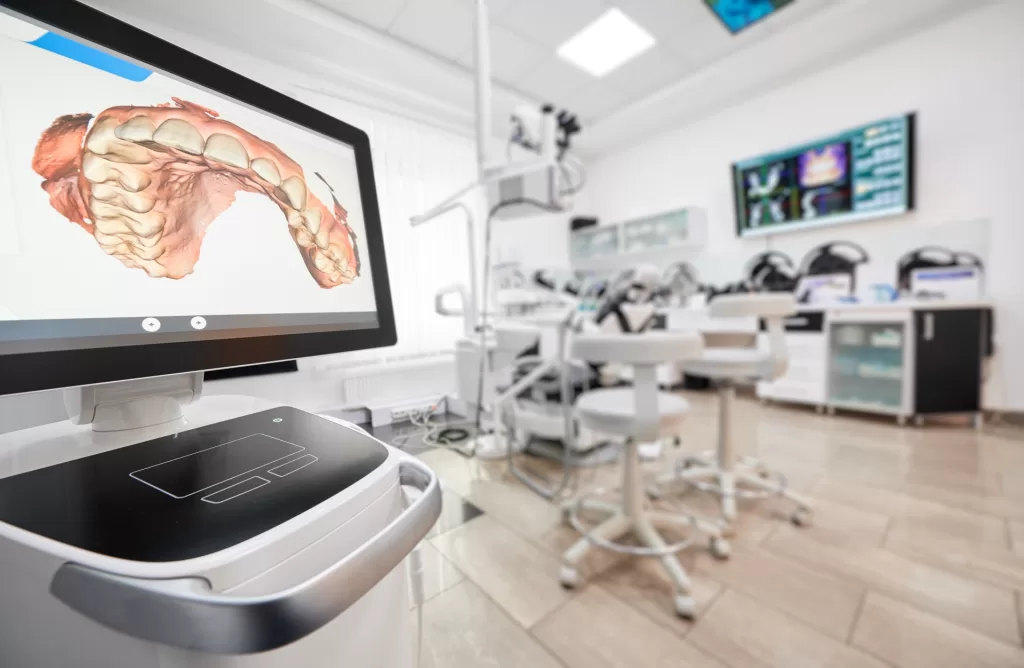
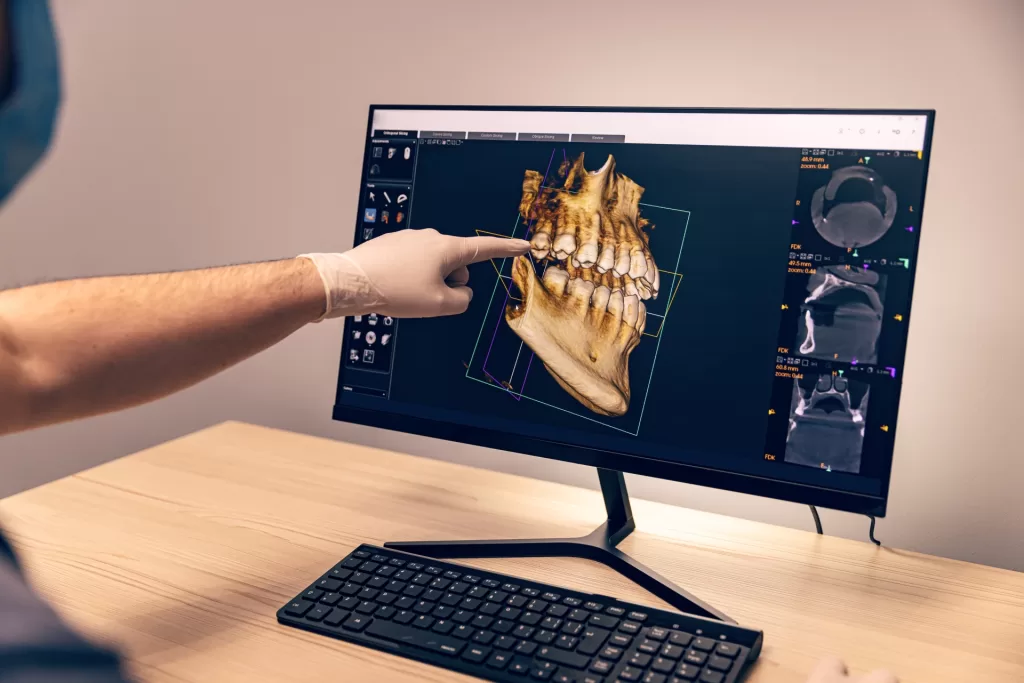
3D imaging in oral surgery refers to advanced imaging procedures, such as Cone Beam Computed Tomography (CBCT), which display detailed three-dimensional representations of the patient´s oral and maxillofacial structures. Unlike traditional 2D imaging, 3D imaging provides a complete view of teeth, bones, nerves, and soft tissues.
This technology captures multiple images from different angles and reconstructs them into a detailed 3D model using specialized software. This 3D model enables the oral surgeon to visualize relevant anatomy in detail, helping them to make an accurate diagnosis, which will also result in proper treatment planning.
At Ridge Oral Surgery, we embrace 3D imaging to give our patients cutting-edge treatment. Let’s understand the key advantages of 3D imaging and how it revolutionizes oral surgery through this blog.
Key Advantages of 3D Imaging in Oral Surgery
From accurate diagnoses to safer, more effective treatment plans, here’s why 3D imaging is a game-changer in oral surgery:
Precision and Accuracy
It gives the oral surgeon a better appreciation of the anatomy and any potential problems. This is especially critical for a procedure like dental implant placement, where the surgeon must fully appreciate bone density and interferences from probable nerve locations to avert complications. 3D imaging improves diagnostic accuracy and enables personalized treatment plans tailored to each patient.
Enhanced Treatment Planning and Reduced Risks
With comprehensive 3D models, we can simulate surgical procedures before performing them, allowing for meticulous planning. This foresight reduces the likelihood of unexpected complications during surgery. For instance, when planning for dental implants, 3D imaging allows us to assess the jawbone’s thickness and health, ensuring optimal implant placement and longevity.

How 3D Imaging Improves Treatment Outcomes
3D imaging not only enhances surgical precision but also leads to better patient outcomes. From increasing the success rate of procedures to improving recovery times, here’s how this technology is making a difference.
Real-Life Examples of Improved Results
Consider a patient requiring a dental implant. Traditional 2D imaging may not reveal bone density issues, increasing the risk of implant failure. But with 3D imaging, we would catch such scenarios before they happen and prepare for bone grafting where necessary, thus increasing the success of that implant. Also, for impacted wisdom teeth, 3D imaging aids us in understanding the exact locations of the impacted teeth with the nerves and other teeth, securing a safe extraction.
Patient Comfort and Faster Recovery Times
Surgical templates enable precise planning, minimizing tissue disruption and reducing post-surgery pain and swelling. Accurately performed, these procedures mean shorter recovery times and a more pleasant experience for our patients. 3D imaging can be advantageous in performing minimally invasive surgeries, consequently lowering the surgical risks, recovery periods, and postoperative discomfort.
Popular Applications of 3D Imaging in Oral Surgery
3D imaging has become an essential tool in modern oral surgery, offering unparalleled precision in diagnosis and treatment planning. Here is how it is used:
Dental Implants: 3D imaging ensures proper assessment of the quality and quantity of bones and requirements for the successful placement of an implant. Precisely calculates the dimensions and location of implants thereby reducing the chances of complications.
Wisdom Teeth Removal: 3D imaging helps assess wisdom teeth placement near nerves and sinuses, ensuring safer extractions. This is the kind of information that 3D imaging is capable of providing for the enhancement of a surgical safety environment.
Bone Grafting: 3D imaging accurately measures bone loss before grafting, improving integration and stability for successful augmentation. Orthognathic Surgery: During orthogenetic surgery, this imaging process shows the jaws’ discrepancy from an anteroposterior as well as a vertical standpoint, leading toward more exact therapeutic movements that will be converted into functional and esthetically improved outcomes.

The Role of 3D Imaging in Preventing Complications
Unlike 2D imaging, 3D scans provide a complete, realistic view of oral structures, helping both patients and surgeons understand the anatomy better. With three-dimensional imaging, we would be able to minimize the risks that come about during procedures that involve complex anatomical areas and reduce the need for follow-up interventions.
Minimizing Risks During Complex Surgeries
3D imaging provides a detailed view of bone structures and nerve pathways, helping to identify potential issues before surgery. The two dimensions in conventional imaging fall short when it comes to the complex anatomy of the oral cavity, which leads to unforeseen challenges appearing in the course of surgery.
The mandibular nerve in dental implantology, for example, should be specifically located, so that nerve damage can be avoided. CBCT imaging gives very accurate mappings of the anatomic landmarks, essential when doing a safe implant procedure. Bone segments can be analyzed by 3D imaging in orthognathic surgery performed on people with jaw deformities to have the exact evaluation of how these segments should align, thus improving the function after surgery.
Reducing the Need for Follow-Up Procedures
Accurate initial surgeries, guided by 3D imaging, markedly reduce the chances of complications demanding further interventions. For instance, the use of bone segments in surgeries affecting the temporomandibular joint (TMJ), with the inaccurate navigation of these bones, would reduce the risk of alignment loss and possibly prevent correction procedures afterward.
Furthermore, in implant dentistry, computer-assisted implant surgery (CAIS) has begun to realize the implementation of planning and placing implants with high accuracy based on 3D imaging. As a result, treatment times are minimized, and the lifespan of implants increases. This, in turn, reduces the risk of complications and the need for further surgeries.
The Future of 3D Imaging in Oral Surgery
The evolution of 3D imaging technology continues to revolutionize oral surgery, with several cutting-edge advancements on the horizon poised to further enhance patient care.
Cutting-edge Advancements on the Horizon
AI-powered 3D imaging enhances early detection and treatment planning by identifying patterns and anomalies in scans. An early example would be bone resorption or pathological changes that can then be treated appropriately.
How 3D Imaging Will Continue to Revolutionize Oral Care
As 3D imaging technology advances, its applications in oral surgery are expected to expand, leading to more personalized and effective treatments. 3D imaging enables precise digital models for custom surgical guides and prosthetics, improving fit and function in dental restorations.
Integration of the two such technologies, that is, 3D imaging and augmented reality, will also transform surgical training and education. For instance, surgeons could perform increasingly complex surgical procedures in virtual reality before performing them on a patient, thus enhancing skill and confidence.

Get Your 3D Imaging From Ridge Oral Surgery
The application of 3-dimensional imaging has changed the entire face of oral surgery in the diagnosis as well as treatment of various dental diseases. 3D imaging provides a highly detailed view of oral structures, leading to more accurate diagnoses, lower risks, and better patient outcomes.
At Ridge Oral Surgery, we use the most up-to-date technologies such as 3D imaging, making sure to the best care standards for its patients concerning treatment. If you think of oral surgery, you can contact us to fix an appointment so you can directly feel the benefits of advanced imaging in treatment planning.
