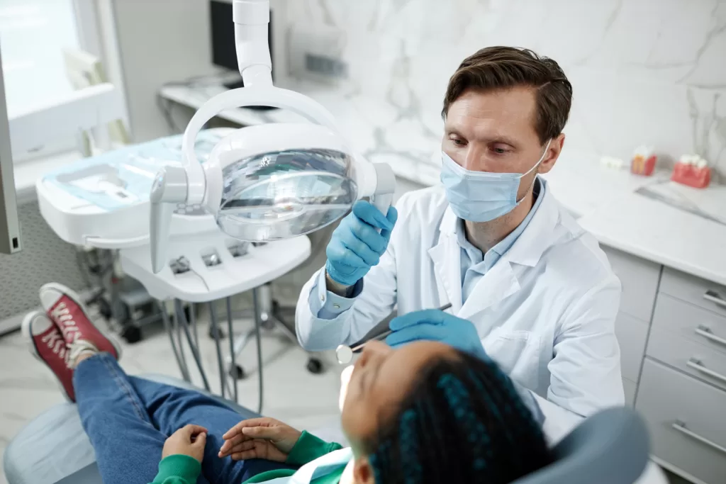
Does the thought of a dental procedure make you anxious? You’re not alone. But thanks to advancements in technology, oral surgery is now more precise, predictable, and less stressful. One game-changer in the industry is 3D imaging, also known as Cone Beam Computed Tomography (CBCT). Unlike traditional X-rays that offer only a flat, two-dimensional view, 3D imaging provides a complete, high-definition picture of your teeth, jaw, and surrounding structures, allowing for better diagnosis and treatment planning.
At Ridge Oral Surgery, we leverage this cutting-edge technology to provide safer, more effective care tailored to each patient. Whether it’s a dental implant, wisdom teeth removal, or a complex procedure, 3D imaging gives us the clarity needed to minimize risks and enhance outcomes. With detailed, real-time visuals guiding every step, you can feel confident knowing your treatment is planned with precision.
This guide explores how 3D imaging is transforming oral surgery, making treatments safer, more precise, and tailored to your needs.
Why 3D Imaging Matters in Oral Surgery
Why is 3D imaging important to oral surgery? Quite simply, it provides a clearer, more defined image of what is occurring under the surface, and this makes every aspect of the procedure more precise and predictable.
A chief benefit is improved diagnostic accuracy. With 3D dental imaging, oral surgeons can observe dental anatomy with more detail than ever before, leaving nothing out. It’s like having a detailed map before going on a journey; you know exactly where you’re going. Another key benefit is reduced radiation exposure, making it a safer alternative to traditional X-rays.
Another revolution is comprehensive treatment planning. The high-level images enable customized treatment plans based on every individual’s own needs. That results in less surprise during surgery and an improved overall outcome.
Minimally invasive procedures are also simpler with 3D imaging. Knowing the region, surgeons can employ smaller cuts, which are less risky, less painful, and quicker to recover from.
Simply put, 3D imaging has revolutionized oral surgery by increasing accuracy, enhancing safety, and making treatment more patient-friendly. It’s a win-win for surgeons and patients alike.
Applications of 3D Imaging at Ridge Oral Surgery
3D imaging revolutionized oral surgery, creating more secure and more accurate procedures. Let’s dissect some of the most important uses and why they are important.
- Dental Implants: Having the implants done correctly requires knowledge of bone shape and nerve location. High-resolution scans provide a precise map to ensure a flawless fit and long-term success.
- Wisdom Teeth Removal: Dealing with impacted wisdom teeth can be difficult. Advanced scans allow doctors to know exactly where the tooth and nerves are, minimizing risks and facilitating smoother removals.
- Bone Grafting: When implants require more bone, advanced imaging evaluates bone quality and plots the graft position for optimal results.
- Tooth Extractions: 3D imaging can help in even the simplest dental extraction by locating roots and surrounding structures and preventing surprises.
- Orthodontics: Straightening teeth is an exact science. High-definition scans provide a complete picture of tooth and jaw placement, allowing for more accurate treatments.
- TMJ Analysis: Advanced imaging is used to provide a more in-depth analysis of jaw pain or movement abnormalities, allowing for more effective treatment.
- Pathology Detection: The sooner cysts, tumors, or other issues are identified, the better. Fine scans can detect these abnormalities rapidly, allowing therapies to be carried out on time.
From diagnosis to therapy, advanced imaging makes oral surgery more efficient, predictable, and personalized for each patient.

3D Imaging vs. Traditional 2D Imaging
Ever noticed that dental procedures feel so much more accurate nowadays? It all lies in the tech behind the scenes. Old-school X-rays show a flat, two-dimensional picture, but the latest scanning technologies allow dentists to view all of it in 3D. It’s like going from a paper map to Google Maps—you have the whole picture.
The clarity is a complete game-changer. Little things that might be easy to miss on regular X-rays are as clear as day, so it’s simpler to catch possible issues early on. And with 3D scans, no more guessing. Overlapping structures that used to merge together are now simple to discern, providing doctors with the entire picture of what’s occurring.
One of the biggest advantages is accuracy. Measurements are much more accurate, so whether it’s placing an implant or planning surgery, everything is planned out with precision. That translates into less risk, less complex procedures, and less time to recover for patients
In short, these scans improve treatments by making them safer, more efficient, and personalized to your specific needs.
What to Expect During Your 3D Imaging Appointment at Ridge Oral Surgery
If you have never had an appointment for 3D imaging, don’t worry. It is easy, fast, and painless. This is what happens when you come in.
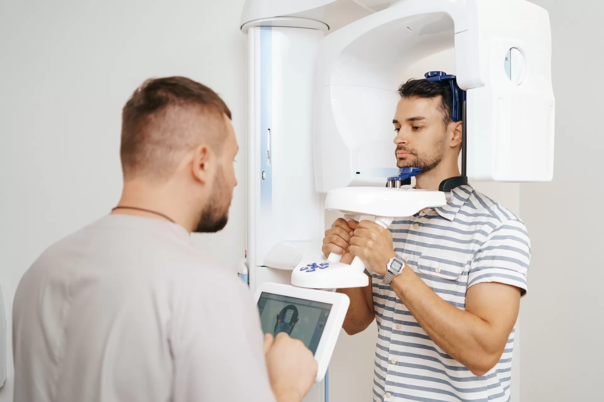
First, we’ll have you take off any jewelry, glasses, or other accessories that may get in the way of the scan. Next, you’ll be escorted to the scanning room, where you’ll either sit or stand. Depending on the type of test. Our experienced staff will position you just right, and then the magic begins.
The scanning process itself takes but a matter of seconds. As the equipment moves slowly about your head, it takes closely detailed pictures of your teeth, jaw, and structures around your face. No discomfort, but merely a time-out while equipment is at work.
After the scan, we will explain the results to you. You will have a clear idea of what is going on under the surface and what treatment could be most suitable for you. We are always happy to answer questions and discuss concerns you may have.
At our office, your care and comfort are our highest concerns. With cutting-edge technology and caring staff, you’re in the best of hands each and every step of the way.

The Future of Oral Surgery is Here: 3D Imaging and Beyond
Oral surgery has progressed significantly, and believe us, it’s amazing the way technology is making everything less stressful and easier. Take 3D imaging, for instance. It presents an extremely clear and detailed image that makes planning treatments much simpler and exacting. Guesswork is over; there is accurate care precisely meant for you.
But that’s not all. We’re employing such things as advanced bone grafting and platelet-rich fibrin (PRF) therapy, which is a fancy name but essentially means applying your own body’s natural growth factors to heal faster. Throw in electronic medical records (EMR) and advanced surgical techniques, and you have an equation that’s designed to make it all safer, quicker, and a whole lot more cozy.
The best part? You’ll feel the difference. From faster procedures to smoother recoveries, these advancements are all about making your experience better. It’s reassuring to know you’re in the hands of a team that’s not just keeping up with technology —we’re leading the way.
Precision, Safety, and Confidence: The Future of Oral Surgery
In the end, it’s all about providing our patients with the optimal care. Adapting technology such as 3D imaging allows us to do just that. It has revolutionized how we perform oral surgery, and everything is now more accurate, efficient, and tailored to their needs. From dental implant planning to the diagnosis of complicated conditions, having that kind of level of detail enables us to make better decisions and provide improved results.
But it’s not all about the technology. It’s about how we leverage it to put patients first. At Ridge Oral Surgery in New Jersey, we’re dedicated to being ahead of the curve, utilizing tools and methods that smooth the way, increase safety, and provide comfort.
If you’ve been delaying a dental procedure or simply want to learn more about how this technology will help you, why not get started? Set up a consultation with our office and see for yourself how 3D imaging can really pay off in your care. We’re here to answer your questions, guide you through the process, and bring you a healthier, happier smile. Let’s take that first step together.
